TAVI-TEC
AI-assisted TAVI planning inside DICOM Vision®
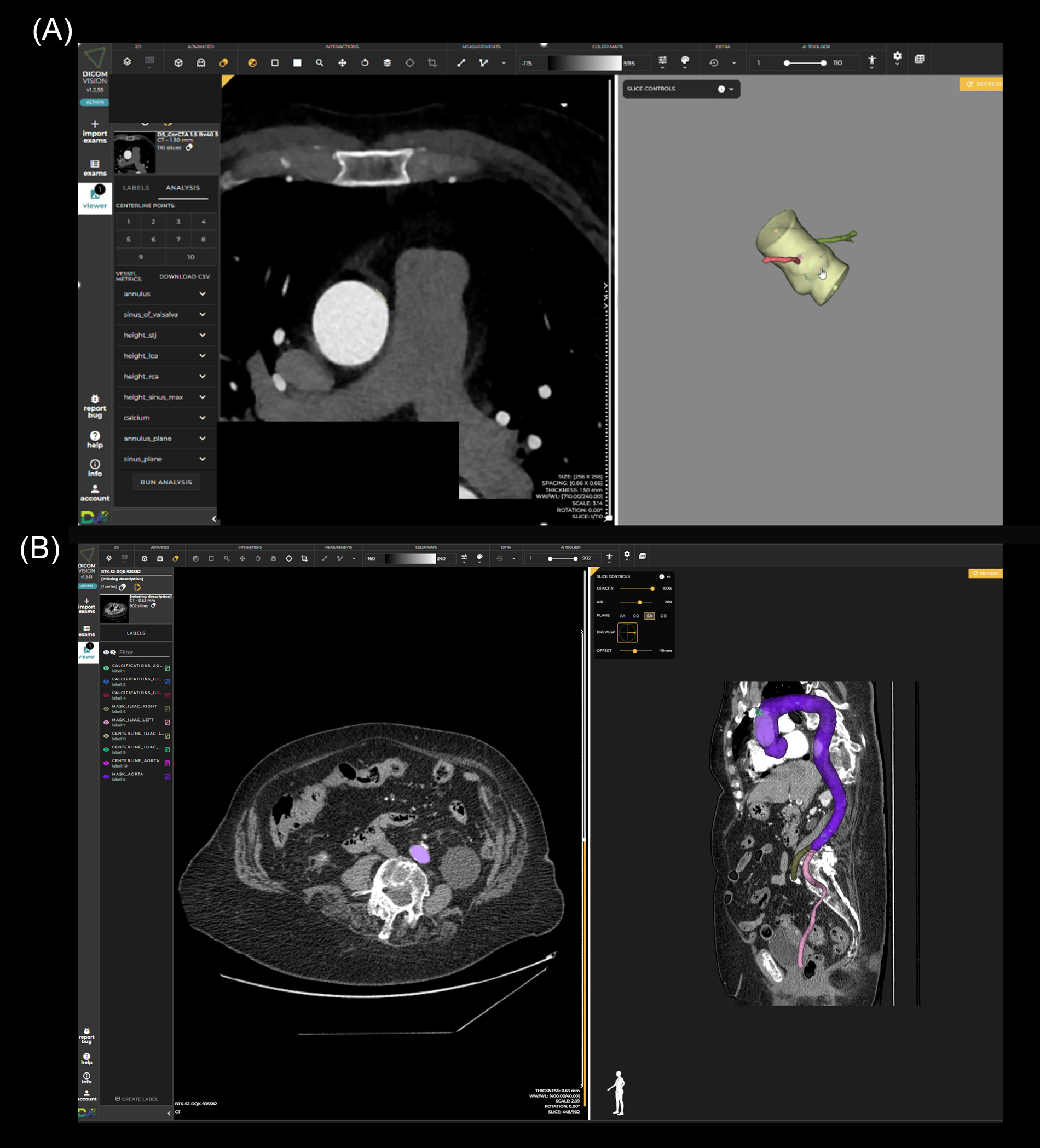
A showcase of how DICOM Vision® brings CTA imaging, AI segmentation, 3D visualization, vascular access assessment and sizing support into a single web-based workflow for TAVI planning.
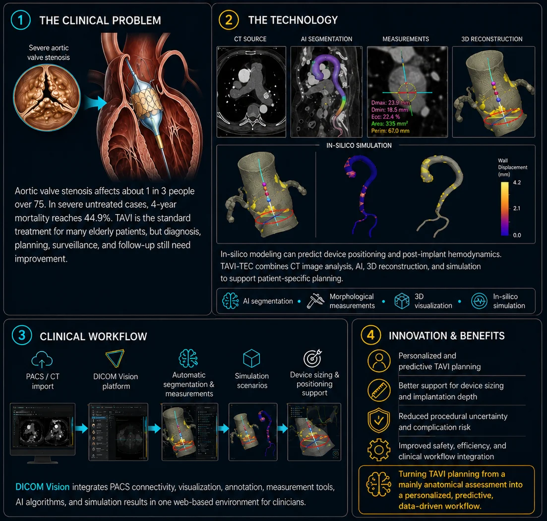
The problem
TAVI planning depends on accurate CTA assessment of the aortic annulus, aortic root, calcifications and vascular access.
Manual measurements can be time-consuming and may vary between operators, especially in borderline cases.
TAVI-TEC explores how AI can help standardize this workflow and bring automatic measurements directly into the clinical imaging environment.
Designed for hospitals, Heart Teams, research groups and medtech projects working on advanced cardiac imaging workflows.
What TAVI-TEC does
TAVI-TEC automatically processes preoperative CTA studies to support:
AI segmentation
Aortic root and vascular structure segmentation.
Automatic measurements
Annular area, perimeter and diameter measurements.
3D anatomical review
Aortic root, calcifications and vascular structures in 3D.
Vascular access assessment
Iliac-femoral evaluation with CPR views and calcification mapping.
Sizing support
Retrospective prosthesis size evaluation for SAPIEN 3 Ultra.
DICOM Vision® integration
Review images, outputs and discussion in one web-based platform.
TAVI-TEC at a glance
From CTA upload to AI output review, the workflow is designed to keep imaging, measurements and discussion inside the same web-based environment.
Video walkthrough
Watch the TAVI-TEC workflow in two short video walkthroughs on YouTube.
CTA-based aortic root analysis
With 3D segmentation and automated measurements. Watch on YouTube.
Full-body CTA review
With segmented aorta and iliac-femoral axis for vascular access assessment. Watch on YouTube.
Why DICOM Vision®
DICOM Vision® provides the web-based environment where clinicians can review the original CTA images, inspect AI-generated outputs, visualize 3D reconstructions, check measurements and discuss the case within a single platform.
The goal is not to replace clinical judgment, but to make advanced imaging analysis easier to access, review and share.
Evaluated on real clinical data
In a retrospective evaluation of 109 TAVI patients, the TAVI-TEC workflow processed CTA studies in 2.2 to 6.4 minutes on a GPU.
The system showed strong agreement with clinician-derived annular measurements (CCC 0.934 for annular area) and achieved 82% agreement with implanted SAPIEN 3 Ultra valve sizes, with most mismatches occurring between adjacent sizes.
Developed with clinical and research partners


TAVI-TEC is the result of a collaboration between clinical teams, academic researchers and medical imaging technology developers.
IRCCS ISMETT contributes clinical expertise and retrospective CTA data from TAVI procedures.
The University of Palermo contributes engineering and bioengineering research expertise.
D/Vision Lab develops the DICOM Vision® web-based imaging platform and supports integration of the TAVI-TEC workflow into the clinical review environment.
The project is supported within the ANTHEM initiative, funded by the European Commission — NextGenerationEU / PNRR.
Build advanced imaging workflows with DICOM Vision®
DICOM Vision® helps clinical and research teams combine DICOM visualization, collaboration, annotation, segmentation and AI-assisted analysis in one web-based platform.
TAVI-TEC is presented as a research and innovation showcase. The workflow is intended to support qualified healthcare professionals and does not replace clinical judgment. Further validation may be required depending on clinical use, deployment context and regulatory status.